Correctly label the anatomical features of the salivary glands – Correctly labeling the anatomical features of the salivary glands is a crucial aspect of research and clinical applications. This comprehensive guide provides a detailed overview of the salivary glands’ anatomy, labeling techniques, and the importance of accurate labeling. By understanding the intricate structure and functions of these glands, we gain valuable insights into their role in maintaining oral health and overall well-being.
The salivary glands are responsible for producing saliva, which plays a vital role in digestion, speech, and protecting the oral cavity from harmful bacteria. Accurate labeling of their anatomical features, including the major (parotid, submandibular, sublingual) and minor salivary glands, enables researchers and clinicians to precisely identify and study their structure, function, and any abnormalities that may arise.
Anatomical Features of Salivary Glands
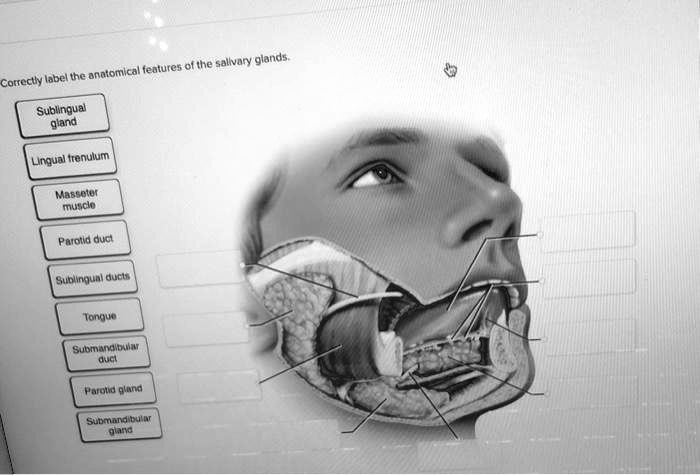
Salivary glands are exocrine glands that secrete saliva into the oral cavity. They play a crucial role in digestion, lubrication, and protection of the oral mucosa. There are three pairs of major salivary glands and numerous minor salivary glands located throughout the oral cavity.
Major Salivary Glands, Correctly label the anatomical features of the salivary glands
* Parotid Gland:The largest salivary gland, located on either side of the face, below the ear. It secretes serous saliva rich in enzymes.
Submandibular Gland
Located below the mandible, it secretes a mixture of serous and mucous saliva.
Sublingual Gland
The smallest salivary gland, located beneath the tongue. It secretes mucous saliva.
Minor Salivary Glands
Minor salivary glands are small glands scattered throughout the oral mucosa. They secrete saliva that lubricates and protects the oral cavity.
Histological Characteristics
Salivary glands are composed of glandular tissue and ducts. The glandular tissue consists of acini, which are the secretory units, and intercalated ducts. The ducts collect saliva from the acini and transport it to the oral cavity.
Labeling Techniques
Accurate labeling of anatomical features is essential for research and clinical applications. Various techniques are used to label salivary glands, including:* Immunohistochemistry:Uses antibodies to bind to specific proteins within the gland, allowing for visualization of specific cell types or structures.
In situ hybridization
Uses complementary DNA probes to bind to specific mRNA sequences, allowing for visualization of gene expression.
Autoradiography
Uses radioactive isotopes to label specific molecules or cells, allowing for visualization of their distribution or activity.
Importance of Correct Labeling: Correctly Label The Anatomical Features Of The Salivary Glands
Accurate labeling of salivary glands is crucial for:* Research:Ensures the accuracy and reproducibility of research findings by providing precise identification of anatomical features.
Clinical Applications
Facilitates accurate diagnosis and treatment of salivary gland diseases by allowing for precise identification of affected areas.
Educational Resources
| Resource | Type | Description |
|---|---|---|
| Salivary Gland Anatomy Course | Online Course | Comprehensive course covering the anatomy, histology, and physiology of salivary glands. |
| Histology of Salivary Glands Textbook | Textbook | Detailed textbook providing in-depth information on the histological structure of salivary glands. |
| Interactive Salivary Gland Simulation | Simulation | Interactive simulation allowing users to explore the anatomy of salivary glands in a virtual environment. |
Advanced Techniques
Emerging techniques for labeling and imaging salivary glands include:* Multiplex Immunofluorescence:Allows for simultaneous labeling of multiple proteins within the gland, providing a more comprehensive view of cell populations and interactions.
Super-Resolution Microscopy
Enhances the resolution of images, allowing for visualization of finer details within the salivary gland.
Confocal Laser Endomicroscopy
Non-invasive technique that allows for in vivo imaging of salivary gland structures in real-time.
FAQ Overview
What are the major salivary glands?
The major salivary glands include the parotid, submandibular, and sublingual glands.
Why is it important to correctly label the anatomical features of the salivary glands?
Correct labeling enables precise identification and study of the glands’ structure, function, and any abnormalities, facilitating research and clinical applications.
What are the different labeling techniques used for salivary glands?
Labeling techniques include histological staining, immunohistochemistry, and molecular imaging, each with its advantages and disadvantages.
