Embark on an educational journey with “The Language of Anatomy Exercise 1,” a comprehensive guide that delves into the fascinating world of anatomical terminology and concepts. As we navigate through this exercise, we’ll unravel the historical evolution of anatomical terms, explore the different anatomical planes and sections, and delve into the complexities of body cavities and regions.
Together, we’ll dissect the intricacies of the skeletal, muscular, and nervous systems, unraveling their structure, function, and classification. We’ll explore the cardiovascular and respiratory systems, understanding the intricate mechanisms that keep us alive. And finally, we’ll delve into the digestive, urinary, and reproductive systems, gaining insights into their vital roles in our overall well-being.
Terminology and Nomenclature
Anatomical terminology has a rich history, evolving over centuries to provide a standardized language for describing the human body. This evolution has been driven by the need for precision and clarity in medical communication and research.
The development of anatomical terminology can be traced back to ancient Greece, where physicians like Hippocrates and Galen made significant contributions. Their work laid the foundation for a systematic approach to describing the body, using terms derived from Greek and Latin.
In the 16th century, the Belgian anatomist Andreas Vesalius published his groundbreaking work “De Humani Corporis Fabrica”, which revolutionized the study of anatomy. Vesalius’s detailed illustrations and descriptions helped establish a more accurate understanding of the human body and its structures.
Over the centuries, anatomical terminology has continued to evolve, with new terms being added and existing terms being refined. In the 19th century, the German anatomist Wilhelm His developed a system of anatomical nomenclature known as the “Nomina Anatomica”, which became the international standard for anatomical terminology.
Importance of Standardized Terminology
The use of standardized anatomical terminology is essential for clear and precise communication among healthcare professionals. It ensures that everyone is using the same terms to refer to the same structures, reducing confusion and errors.
Standardized terminology also facilitates the sharing of medical information across different languages and cultures. It allows researchers and clinicians from different parts of the world to collaborate and exchange knowledge without having to worry about language barriers.
Additionally, standardized terminology is important for teaching and learning anatomy. It provides a common language that students and educators can use to communicate complex anatomical concepts.
Anatomical Planes and Sections
Anatomical planes and sections are essential tools for visualizing and understanding the human body. They provide a framework for describing the location and orientation of anatomical structures.
Anatomical Planes
The three primary anatomical planes are:
- Sagittal plane:Divides the body into left and right halves.
- Frontal (coronal) plane:Divides the body into anterior (front) and posterior (back) halves.
- Transverse (horizontal) plane:Divides the body into superior (upper) and inferior (lower) halves.
Anatomical Sections
Anatomical sections are used to create two-dimensional views of three-dimensional structures. There are three main types of anatomical sections:
- Cross-section:Cut perpendicular to the long axis of the body or a body part.
- Longitudinal section:Cut parallel to the long axis of the body or a body part.
- Oblique section:Cut at an angle to the long axis of the body or a body part.
The type of section used depends on the specific information that is needed. For example, a cross-section is often used to visualize the internal structure of an organ, while a longitudinal section is often used to visualize the relationship between different structures along the length of the body.
Comparison of Anatomical Sections
| Type of Section | Orientation | Uses |
|---|---|---|
| Cross-section | Perpendicular to the long axis | Visualize internal structure of organs |
| Longitudinal section | Parallel to the long axis | Visualize relationship between structures along the length of the body |
| Oblique section | At an angle to the long axis | Visualize structures that are not aligned with the other planes |
Body Cavities and Regions
The human body consists of various cavities and regions, each with specific functions and contents. These cavities provide protection, support, and organization to the internal organs.
Major Body Cavities
The major body cavities include:
Cranial Cavity
Contains the brain and its protective structures.
Vertebral Canal
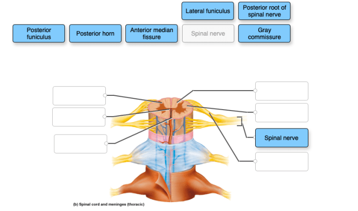
Extends within the vertebral column, housing the spinal cord and nerve roots.
Thoracic Cavity
Enclosed by the ribs and diaphragm, it contains the heart, lungs, and major blood vessels.
Abdominal Cavity
Located below the diaphragm, it houses the stomach, intestines, liver, pancreas, and other digestive organs.
Pelvic Cavity
Situated within the pelvis, it contains the reproductive organs, bladder, and rectum.
Subdivisions of the Abdominal Cavity
The abdominal cavity is further subdivided into four quadrants and nine regions for clinical purposes:
Quadrants
Right Upper Quadrant (RUQ)
Left Upper Quadrant (LUQ)
Right Lower Quadrant (RLQ)
Left Lower Quadrant (LLQ)
Regions
Epigastric
Umbilical
Hypogastric
Right Hypochondriac
Left Hypochondriac
Right Lumbar
Left Lumbar
Right Iliac
Left Iliac
These subdivisions aid in locating and describing the position of organs within the abdomen during physical examinations and diagnostic procedures.
Diagram of Body Cavities
[Diagram showing the relationships between the major body cavities, including the cranial cavity, vertebral canal, thoracic cavity, abdominal cavity, and pelvic cavity.]
Skeletal System
The skeletal system is the framework of the body, providing support, protection, and movement. It consists of bones, joints, and cartilage, which work together to allow for a wide range of functions.
Structure and Function of Bones
Bones are hard, mineralized tissues that form the majority of the skeletal system. They provide structural support for the body, protect vital organs, and facilitate movement. Bones also store minerals, such as calcium and phosphorus, and produce blood cells.
Types of Bones
There are five main types of bones, each with a specific structure and function:
- Long bones: Found in the limbs, they are longer than they are wide and provide support and movement.
- Short bones: Found in the wrists and ankles, they are cube-shaped and provide stability.
- Flat bones: Found in the skull and rib cage, they are thin and broad, providing protection and support.
- Irregular bones: Found in the spine and face, they have complex shapes and provide support and protection.
- Sesamoid bones: Small, round bones embedded in tendons, they protect tendons from excessive friction.
Joints
Joints are the points of contact between two or more bones. They allow for movement and provide stability to the body. There are several types of joints, each with a different range of motion:
- Synarthroses: Immovable joints, such as those found in the skull.
- Amphiarthroses: Slightly movable joints, such as those found between the vertebrae.
- Diarthroses: Freely movable joints, such as those found in the knees and elbows.
Muscular System
The muscular system is responsible for movement, posture, and heat production. Muscles are composed of specialized cells called muscle fibers, which are capable of contracting and relaxing.The three main types of muscles are skeletal muscle, smooth muscle, and cardiac muscle.
Skeletal muscles are attached to bones and are responsible for voluntary movement. Smooth muscles are found in the walls of organs and blood vessels and are responsible for involuntary movements such as digestion and blood flow. Cardiac muscle is found only in the heart and is responsible for pumping blood.
Major Muscle Groups and Functions
The following table summarizes the major muscle groups and their functions:| Muscle Group | Function ||—|—|| Axial Muscles | Support and move the head, neck, and trunk || Appendicular Muscles | Move the limbs || Respiratory Muscles | Involved in breathing || Digestive Muscles | Involved in digestion || Urinary Muscles | Involved in urination || Reproductive Muscles | Involved in reproduction |
Nervous System
The nervous system is the control center of the body, responsible for coordinating actions and reactions, processing information, and maintaining homeostasis. It comprises a complex network of specialized cells that transmit electrical and chemical signals throughout the body.The nervous system can be divided into two main divisions: the central nervous system (CNS) and the peripheral nervous system (PNS).
The CNS consists of the brain and spinal cord, which act as the central processing unit and command center. The PNS comprises all the nerves that extend from the CNS to the rest of the body, serving as communication pathways between the CNS and the organs, muscles, and sensory receptors.
Major Components of the Brain and Spinal Cord
Brain:The brain is the primary control center of the nervous system, responsible for higher-level functions such as consciousness, thought, memory, and emotion. It is divided into three main parts: the cerebrum, cerebellum, and brainstem. Cerebrum:The cerebrum is the largest part of the brain, responsible for cognitive functions, including reasoning, problem-solving, language, and memory.
Cerebellum:The cerebellum is located at the back of the brain and is responsible for coordination, balance, and motor control. Brainstem:The brainstem connects the cerebrum and cerebellum to the spinal cord. It controls essential life functions such as breathing, heart rate, and digestion.
Spinal Cord:The spinal cord is a long, cylindrical bundle of nerve fibers that extends from the brainstem down the back. It serves as a communication pathway between the brain and the rest of the body, transmitting sensory and motor signals.
Cardiovascular System
The cardiovascular system is a complex network of organs and vessels that work together to pump blood throughout the body. The heart is the central organ of the cardiovascular system, and it is responsible for pumping blood through the body’s arteries, veins, and capillaries.
The circulatory system is responsible for transporting oxygen, nutrients, hormones, and other essential substances to the body’s cells, and for removing waste products from the cells.
Structure and Function of the Heart
The heart is a muscular organ that is located in the chest cavity. It is divided into four chambers: two atria (upper chambers) and two ventricles (lower chambers). The atria receive blood from the body, and the ventricles pump blood out to the body.
The heart valves prevent blood from flowing backward through the heart.
Circulation of Blood through the Body
The heart pumps blood through the body in two circuits: the pulmonary circulation and the systemic circulation. The pulmonary circulation is the circuit that carries blood from the heart to the lungs and back. The systemic circulation is the circuit that carries blood from the heart to the rest of the body and back.
Major Blood Vessels and Their Functions
The major blood vessels in the body are the arteries, veins, and capillaries. Arteries carry blood away from the heart, veins carry blood back to the heart, and capillaries are small vessels that allow oxygen and nutrients to pass from the blood into the body’s cells.
- Arteriescarry oxygenated blood away from the heart to the rest of the body.
- Veinscarry deoxygenated blood back to the heart from the rest of the body.
- Capillariesare small blood vessels that allow oxygen and nutrients to pass from the blood into the body’s cells.
Respiratory System: The Language Of Anatomy Exercise 1
The respiratory system is responsible for taking in oxygen and expelling carbon dioxide. It consists of the lungs, airways, and respiratory muscles. The lungs are two large, spongy organs located in the chest cavity. The airways are a series of tubes that carry air to and from the lungs.
The respiratory muscles are responsible for expanding and contracting the lungs, which creates the airflow necessary for breathing.
The process of respiration begins when we inhale. Air enters the nose or mouth and travels through the pharynx (throat) and larynx (voice box). It then enters the trachea (windpipe), which branches into two bronchi. The bronchi enter the lungs and divide into smaller and smaller airways called bronchioles.
The bronchioles end in tiny air sacs called alveoli. The alveoli are surrounded by capillaries, which are tiny blood vessels. Oxygen from the air in the alveoli diffuses across the capillary walls and into the bloodstream. Carbon dioxide from the bloodstream diffuses across the capillary walls and into the alveoli.
The carbon dioxide is then exhaled through the airways and out of the body.
Major Organs of the Respiratory System
The major organs of the respiratory system include:
- Lungs: The lungs are the primary organs of respiration. They are responsible for exchanging oxygen and carbon dioxide between the bloodstream and the air.
- Airways: The airways are a series of tubes that carry air to and from the lungs. The main airways are the trachea, bronchi, and bronchioles.
- Respiratory muscles: The respiratory muscles are responsible for expanding and contracting the lungs, which creates the airflow necessary for breathing.
Digestive System
The digestive system is responsible for the breakdown of food into nutrients that can be absorbed and used by the body. It consists of a series of organs that work together to break down food, absorb nutrients, and eliminate waste products.The
process of digestion begins in the mouth, where food is chewed and mixed with saliva. Saliva contains enzymes that begin to break down carbohydrates. The food is then swallowed and travels down the esophagus to the stomach. The stomach secretes gastric juices, which contain hydrochloric acid and enzymes that further break down food.
The food is churned and mixed in the stomach, forming a semi-liquid substance called chyme.The chyme then passes into the small intestine, where it is further broken down by enzymes from the pancreas and bile from the liver. The nutrients from the food are absorbed into the bloodstream through the walls of the small intestine.
The remaining waste products pass into the large intestine, where water is absorbed and the waste is formed into feces. The feces are then eliminated from the body through the rectum.
Major Organs of the Digestive System
The major organs of the digestive system include:
- Mouth
- Esophagus
- Stomach
- Small intestine
- Large intestine
- Pancreas
- Liver
- Gallbladder
Each of these organs plays a specific role in the process of digestion. The mouth breaks down food into smaller pieces and mixes it with saliva. The esophagus transports food from the mouth to the stomach. The stomach secretes gastric juices that break down food and churn it into a semi-liquid substance.
The small intestine absorbs nutrients from food. The large intestine absorbs water from food and forms feces. The pancreas secretes enzymes that break down food. The liver produces bile, which helps to break down fats. The gallbladder stores bile.
Urinary System
The urinary system plays a crucial role in maintaining the body’s fluid balance, eliminating waste products, and regulating blood pressure. It comprises several organs that work together to filter and excrete urine.The process of urine formation begins in the kidneys, where blood is filtered to remove waste products and excess water.
The filtered fluid, known as filtrate, then flows through the renal tubules, where essential substances are reabsorbed into the bloodstream. The remaining fluid, which contains waste products, forms urine. Urine is then transported through the ureters to the bladder, where it is stored until it is expelled through the urethra during urination.
Major Organs of the Urinary System, The language of anatomy exercise 1
- Kidneys:The primary organs responsible for filtering blood and producing urine.
- Ureters:Tubes that carry urine from the kidneys to the bladder.
- Bladder:A muscular organ that stores urine until it is released.
- Urethra:A tube that transports urine from the bladder to the outside of the body.
The urinary system is a vital part of the body’s overall health and well-being. It helps maintain fluid balance, eliminate waste products, and regulate blood pressure, ensuring proper bodily functions.
FAQs
What is the importance of using standardized terminology in anatomy?
Standardized terminology ensures consistency and clarity in communication among healthcare professionals. It eliminates confusion and misinterpretation, facilitating accurate diagnosis, treatment, and research.
How many primary anatomical planes are there?
There are three primary anatomical planes: sagittal, coronal, and transverse.
What is the function of the skeletal system?
The skeletal system provides support, protection, movement, and storage of minerals.